- About
-
Solutions
-
Services
-
Biosciences
- PureDisc™ Enabled Membrane Protein Production
- Assay Development and Transfer
- Biochemistry / Enzymology
- Biomarker Services
- Biophysics
- Cell-Based Assays
- Compound screening and cascade design
- Cryo-EM
- Fibrosis Assays
- Project Management and Consultancy Services
- Protein Expression
- Structural Biology
- X-ray Crystallography
- Chemistry
- Integrated Drug Discovery
- Computer Aided Drug Design
- Hit ID and Screening Services
- DMPK Services
-
Biosciences
- Target Classes and Modalities
- Therapeutic Areas
-
A-Z
- A
- B
- C
- D
- E
- F
- G
- H
- I
- K
- L
- M
- N
- O
- P
- R
- S
- T
- V
- X
-
Services
- Library
- News & Events
- Careers
- PureDisc™
Cryogenic Electron Microscopy (Cryo-EM)
deep expertise • flexible solutions • close collaboration • rapid delivery
Cryo-electron microscopy (cryo-EM) has advanced rapidly in recent years, driven by innovations in detector technology, image processing and sample preparation. These developments now enable high-resolution structure determination with increasingly rapid turnaround, transforming cryo-EM into a powerful tool for structure-based drug discovery (SBDD). In particular, cryo-EM has opened access to challenging targets that are often intractable by X-ray crystallography, including membrane proteins, large multi-protein assemblies, transient complexes, and highly dynamic or heterogeneous systems.
At Domainex, cryo-EM is fully integrated within our core capabilities. Combined with our expertise in protein science, biophysics, computational chemistry (CADD), and medicinal chemistry, we deliver a cohesive approach that accelerates and de-risks your drug discovery programmes.
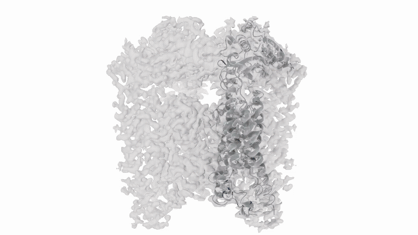
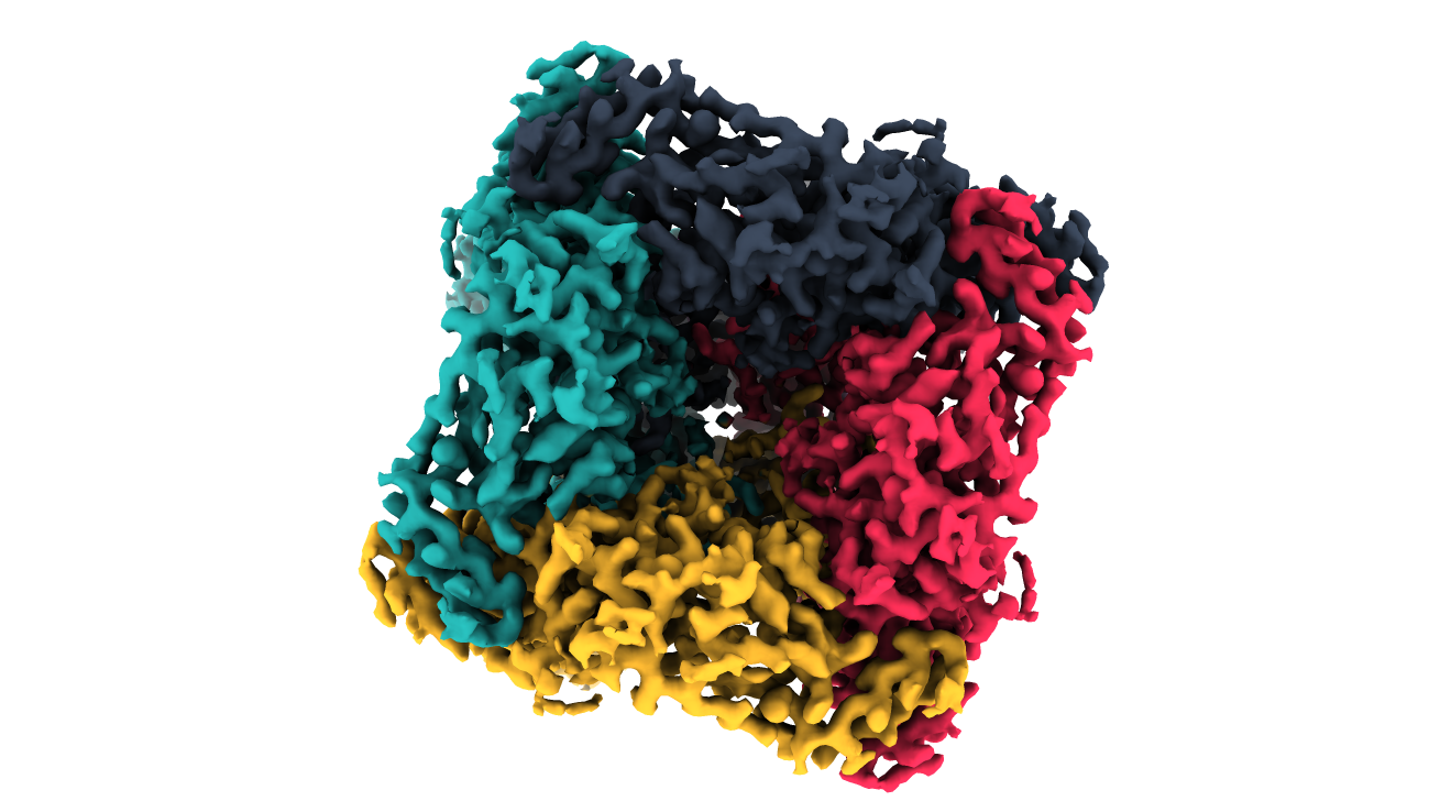
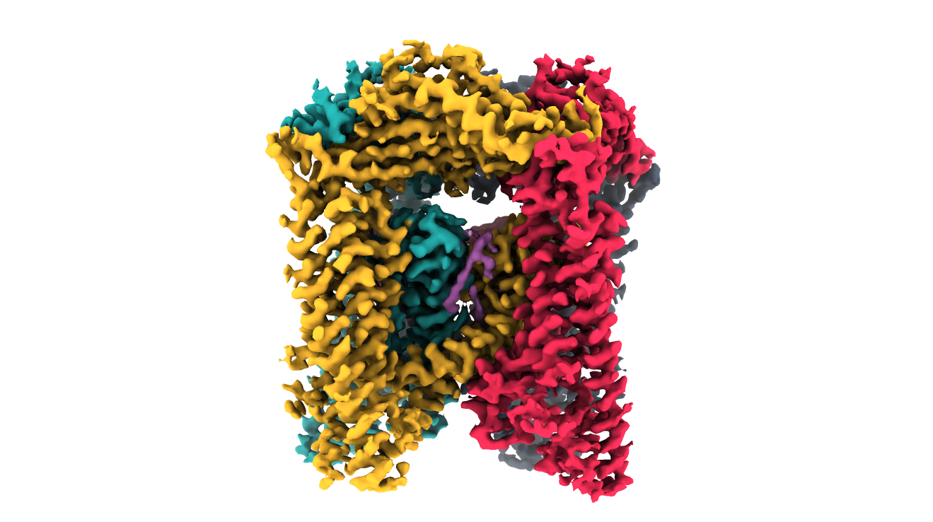
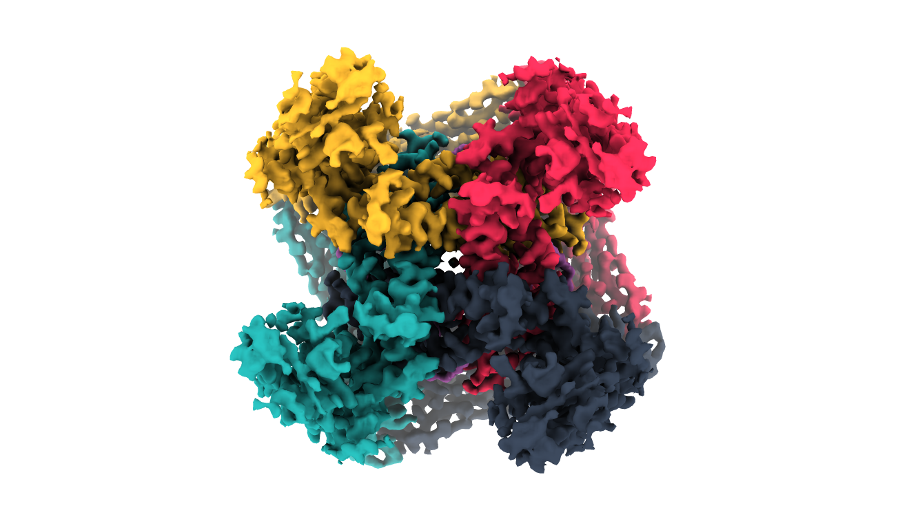
Figure 1. a. Animation of TRPML3 ion channel isolated using Domainex's PureDiscTM technology and b. Views of the PureDisc-isolated TRPML3 ion channel from the top (left), side (middle) and bottom (right)
Key Advantages for Drug Discovery
At Domainex, Cryo‑EM is an essential platform in drug discovery especially for targets >100 kDa that are not always well suited to X-ray crystallography such as:
- Membrane proteins - see TRPML3 case-study and our PureDiscTM-enabled membrane protein production.
- Ternary protein complexes such as those formed by molecular glue or bivalent degrader molecules
- Proteins with non-heterogeneous post translational modifications such as N-linked glycosylation
- Dynamic proteins or those containing disordered regions
- Any protein target >100 kDa
Fully Integrated Cryo‑EM Workflow
Our platform delivers a seamless, end‑to‑end workflow designed to maximise success and accelerate timelines:
Week 1
Week 2
Week 3
Week 4
Week 5
Week 1 — Sample Preparation
- Protein quality assessment and optimisation
- Sample stabilisation strategies, including ligands, nanobodies, and complex formation
- Grid preparation using rapid vitrification technologies
Week 2 — Grid Screening and Data Collection
- Access to state-of-the-art electron microscopes and direct electron detectors
- Grid screening and iterative optimisation of particle distribution, concentration, and orientation
- On-the-fly pre-processing and 2D classification of multi-grid screening datasets to guide and optimise data collection strategies
- High-throughput acquisition of high-resolution datasets
Week 3 — Initial Map Generation
- Automated image pre-processing, particle picking, and classification
- Generation of initial 3D reconstructions • Structural heterogeneity assessment through 3D classification
Week 4 — High-Resolution Map Refinement
- Advanced 3D reconstruction and refinement workflows
- Generation of high-resolution density maps suitable for atomic model building
- Comprehensive map quality assessment and validation
Week 5 — Structure Determination, Model Building and Refinement
- Atomic model building, refinement, and validation
- Detailed analysis of protein–ligand and protein–protein interactions
- Support for structure-based drug design (SBDD) and project decision-making
- Delivery of publication-ready structures, maps, and comprehensive reports
|
|
|
|
|
Outcome: High-quality structural insights delivered through an integrated workflow, enabling confident biological interpretation and informed project decisions.
Our team of highly experienced structural biologists bring deep expertise in applying structural biology to drug discovery, helping to drive informed decision-making and accelerate programme progression.
Frequently Asked Questions
1. What is cryo-electron microscopy (cryo-EM) and how does it contribute to structure-based drug design?
Cryo-electron microscopy (Cryo-EM) has become a powerful tool for structure-based drug discovery (SBDD), providing high-resolution structural information for targets that can be difficult to study using traditional X-ray crystallography approaches.
By enabling structural insights into challenging biological systems, Cryo-EM can help accelerate hit identification, lead optimisation and mechanism-of-action studies.
2. For which target classes is cryo-EM particularly advantageous?
Cryo-EM can be used on any soluble protein >100kDa and is especially well suited to structurally complex or intractable targets, including:
- Membrane proteins and ion channels
- Large macromolecular assemblies and multi-protein complexes
- Ternary complexes (e.g. PROTACs, molecular glues)
- Conformationally dynamic or heterogeneous systems
- Proteins with significant post-translational modifications (e.g. glycosylation)
Domainex scientists can assess your target and advise whether Cryo-EM is the most appropriate structural biology approach for your programme.
3. How is cryo-EM integrated within Domainex’s discovery platform?
At Domainex, cryo-EM is embedded within a multidisciplinary discovery framework that tightly integrates:
- Protein science: Construct design, expression system optimisation, purification, and stabilisation strategies
- Biophysics: Orthogonal validation of protein quality, conformational integrity, and ligand engagement
- Computational chemistry (CADD): Structural modelling, binding mode elucidation, and compound design
- Medicinal chemistry: Structural biology uncovers the 3D shapes of disease-causing proteins, allowing medicinal chemists to custom-design small molecules or biologics that bind precisely to these targets to neutralise them
This unified approach enables iterative optimisation cycles between experimental and computational disciplines, accelerating decision-making and reducing programme risk.
4. How does protein science underpin cryo-EM success?
High-quality, conformationally homogeneous protein samples are critical determinants of cryo-EM success. Domainex applies advanced protein science strategies including:
- Rational construct engineering to remove disorder and enhance stability
- Optimisation of expression systems (e.g. mammalian, insect, and bacterial)
- Use of ligands, cofactors, antibodies or nanobodies to stabilise specific conformations
- Complex reconstitution for multi-component systems
These approaches maximise particle quality, improve grid behaviour, and increase the likelihood of obtaining high-resolution reconstructions.
5. What is the role of biophysics in supporting cryo-EM workflows?
Biophysical characterisation provides essential, quantitative validation of both protein samples and ligand interactions prior to and alongside cryo-EM studies. Techniques may include:
- Differential Scanning Fluorimetry (DSF) and calorimetry for stability assessment
- Grating-Coupled Interferometry and Surface Plasmon Resonance (GCI/SPR) or Spectral Shift for binding kinetics and affinity
- Dynamic Light Scattering (DLS) and mass photometry for oligomeric state and homogeneity
Integration of these datasets ensures that cryo-EM structures are interpreted within a robust physicochemical context, enhancing the reliability of downstream SBDD decisions.
6. How does CADD enhance cryo-EM-derived structural insights?
Domainex use Computational chemistry (CADD) to extract maximum value from cryo-EM datasets through:
- Atomic model refinement and validation against density maps
- Binding pose prediction and optimisation
- Structure-based virtual screening and hit identification
- Free energy and molecular dynamics simulations to explore conformational landscapes
The iterative integration of experimental structures with computational modelling enables rapid progression from structural insight to chemical optimisation.
7. What are the key stages of the Domainex cryo-EM workflow?
Domainex delivers a comprehensive, end-to-end cryo-EM workflow comprising:
- Sample preparation - protein optimisation, complex formation, and vitrification
- Grid screening and optimisation - iterative refinement of particle distribution and orientation
- Data acquisition - high-throughput collection using state-of-the-art instrumentation
- Image processing - particle picking, 2D classification, and 3D reconstruction
- Map refinement - high-resolution density map generation and validation
- Model building - atomic interpretation and structural analysis
Each stage is tightly integrated with upstream and downstream scientific disciplines to maximise efficiency and data quality.
8. What outputs and deliverables can be expected?
Cryo-EM projects at Domainex generate:
- High-resolution 3D density maps
- Refined, validated atomic models
- Detailed analyses of protein-ligand or protein-protein interfaces
- Mechanistic insights into target biology and modulation
- Comprehensive reports supporting programme progression
All deliverables are aligned to enable direct application in medicinal chemistry and SBDD workflows.
9. Can Domainex use cryo-EM to support ligand-binding and complex mechanism studies?
Yes. Cryo-EM is increasingly applied to resolve ligand-bound states and higher-order complexes, including:
- Small molecule binding (where resolution permits)
- Ternary complexes relevant to targeted protein degradation
- Allosteric modulation and conformational transitions
When complemented by biophysical validation and CADD analysis, cryo-EM enables detailed mechanistic understanding of target engagement and modulation.
10. How does Domainex accelerate timelines and mitigate technical risk?
Domainex’s co-located expertise across structural biology, protein science, biophysics, and computational chemistry enables:
- Rapid experimental iteration across disciplines
- Early identification and mitigation of developability risks
- Data-driven progression through discovery milestones
- Efficient translation of structural insights into chemical matter
This integrated model reduces reliance on fragmented workflows and provides a coherent, high-confidence path from target validation to candidate optimisation.
Start your next project with Domainex
Contact one of our experts today